Calendula officinalis (flower)

From AHPA Botanical Identity References Compendium
Revision as of 21:15, 15 March 2014 by Staffer (Talk | contribs)

Jump to: navigation, search


Contents

Nomenclature

Calendula officinalis L.   Asteraceae  
Standardized common name (English): calendula

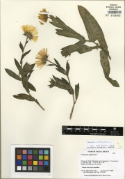
Botanical Voucher Specimen

Calendula officinalis - Tropicos.org (López López, Laura Margarita - 0144) 

Reference see Tropicos.org. Missouri Botanical Garden. 17 Oct 2013 <http://www.tropicos.org/Image/100229512>, Tropicos.org. Missouri Botanical Garden. 17 Oct 2013 <http://www.tropicos.org/Image/89040>, Missouri Botanical Garden. 29 Oct 2013 <http://www.tropicos.org/Image/83485>.

Source: MOBOT, Tropicos.org [1]

TropicosLogo.gif
(thumbnail)
Calendula officinalis - Tropicos.org (López López, Laura Margarita - 0144)

Macroscopic Characteristics

Microscopic Characteristics

The dried ligulate florets of Calendula officinalis Linne.

The N. F. drug is described as in "florets from 15 to 25 mm. in length, yellow- or orange-colored, one to three-toothed, four- to five-veined, margin nearly entire, the short hairy tube occasionally enclosing the remnants of a filiform style and bifid stigma.

The powdered drug is light yellow to orange-yellow and, when examined under the microscope, exhibits a few characteristic, non-glandular hairs, consisting of a double row of thin-walled, more or less collapsed cells, with a one- or two-celled summit, and up to about 0.8 mm. in length; elongated epidermal cells with thin, somewhat wavy walls, a striated surface, and containing irregular chromo-plasts and oil-like globules, the latter coalescing when mounted in hydrated chloral T.S.; pollen grains, more or less spherical, with numerous spiniae projections, three pored, and up to 0.04 mm. in diameter; tracheae about 0.009 mm. in width with spiral and annular markings; prisms or rosette aggregates of calcium oxalate from 0.002 to 0.004 mm. in diameter." N.F.

The odor is much stronger in the fresh than in the dry flowers, and on exposure to light, the orange-red or yellow color fades.

Source: United States Dispensatory (1918) [2]

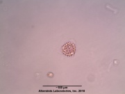
Large spherical pollen grain showing sharp spiny exine with three pores from Calendula officinalis flower viewed at 400x with Acidified Chloral Hydrate Glycerol Solution.cellular structures identified in Calendula officinalis flower are the large spherical pollen grain showing sharp spiny exine with three pores and a fibrous layer of anther in surface view when observed at 400x with Acidified Chloral Hydrate Glycerol Solution.

Source: Elan M. Sudberg, Alkemist Laboratories [3]

AP-LOGO-Laboratories Crop - Copy.jpg
Calendula officinalis L. -Asteraceae--1.jpg


High Performance Thin Layer Chromatographic Identification

AP-LOGO-Laboratories Crop - Copy.jpg
(thumbnail)
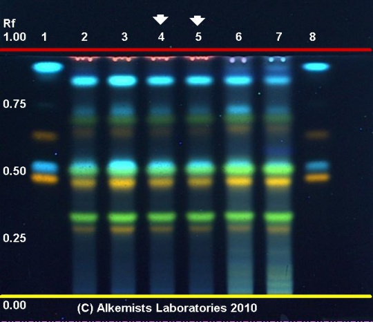
Calendula officinalis HPTLC ID - Natural Product Reagent + PEG UV 365 nm

Marigold (flower) (Calendula officinalis)

Lane Assignments Lanes, from left to right (Track, Volume, Sample):

  1. 1 μL Rutin, Caffeic Acid, Hyperoside, Chlorogenic Acid ~ 0.1% in Methanol
  2. 3 μL Calendula officinalis-1 (flower)
  3. 3 μL Calendula officinalis-2 (flower)
  4. 3 μL Calendula officinalis-3 (flower)
  5. 3 μL Calendula officinalis-3 (flower)
  6. 3 μL Calendula officinalis-4 (flower)
  7. 3 μL Calendula officinalis-5 (flower)
  8. 1 μL Rutin, Caffeic Acid, Hyperoside, Chlorogenic Acid ~ 0.1% in Methanol

Reference materials used here have been authenticated by macroscopic, microscopic &/or TLC studies according to the reference source cited below held at Alkemists Laboratories, Costa Mesa, CA. 

Stationary Phase Silica gel 60, F254, 10 x 10 cm HPTLC plates 

Mobile Phase ethyl acetate: HCOOH: Acetic acid: H2O [10/1.1/1.1/2.4] 

Sample Preparation Method 0.3 g + 3 ml CH3OH sonicate/heat @ 50° C ~ 1/2 hr. 

Detection Method Natural Product Reagent + PEG -> UV 365 nm 

Reference see Plant Drug Analysis, Wagner, H., 1996


Source: Elan M. Sudberg, Alkemist Laboratories [4]

HPTLC-assoc-Logo-farbig-Text-schwarz-300x47.png
(thumbnail)
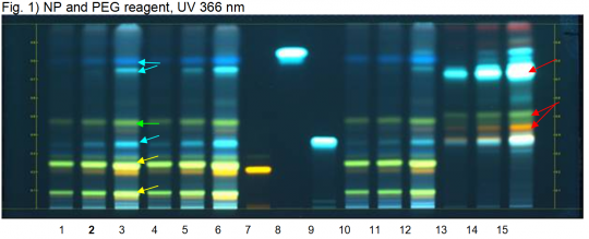
Calendula officinalis (flower) HPTLC ID - NP and PEG reagent, UV 366 nm

Calendula (flower) (Calendula officinalis)

Lane Assignments Lanes, from left to right (Track, Volume, Sample):

  1. 3 µL Calendula flower
  2. 6 µL Calendula flower
  3. 12 µL Calendula flower
  4. 1 µL Calendula flower (Ph. Eur. extraction)
  5. 2 µL Calendula flower (Ph. Eur. extraction)
  6. 4 µL Calendula flower (Ph. Eur. extraction)
  7. 2 µL Rutin
  8. 2 µL Caffeic acid
  9. 2 µL Chlorogenic acid
  10. 4 µL Calendula flower (old)
  11. 8 µL Calendula flower (old)
  12. 16 µL Calendula flower (old)
  13. 2 µL Arnica flower
  14. 4 µL Arnica flower
  15. 8 µL Arnica flower 

Reference Sample(s) Reference: Dissolve 2.5 mg of rutin in 5 mL of methanol. Dissolve 3 mg of caffeic acid in 5 mL of methanol. Optional: dissolve 1.5 mg of chlorogenic acid in 5 mL of methanol. 

Stationary Phase Stationary phase, i.e. Silica gel 60, F254 

Mobile Phase Ethyl acetate, formic acid, water 80:10:10 (v/v/v) 

Sample Preparation Method Sample: Mix 0.5 g of powdered sample with 5 mL of methanol and sonicate for 10 minutes, then centrifuge or filter the solutions and use the supernatants / filtrates as test solutions.

Derivatization reagent: 1.) NP reagent Preparation: 1 g of natural products reagent in 200 mL of ethyl acetate 2.) PEG reagent Preparation: 10 g of polyethylene glycol 400 in 200 mL of dichloromethane Use: Heat plate for 3 min at 100°C, dip (time 0, speed 5) in NP reagent while still hot, dry and dip (time 0, speed 5) in PEG reagent 

Detection Method Saturated chamber; Developing distance 70 mm from lower edge; Relative humidity 33% 

Other Notes Images presented in this entry are examples and are not intended to be used as basis for setting specifications for quality control purposes. System suitability test: Rutin: a yellow fluorescent zone at Rf ~ 0.21; Caffeic acid: a white-blue fluorescent zone at Rf ~ 0.85

Identification: Compare result with reference images. The fingerprint of the test solution is similar to that of the corresponding botanical reference sample. Additional weak zones may be present. The chromatogram of the test solution shows a yellow zone corresponding to the reference material rutin. Right above and below rutin there are a green zone each at Rf ~ 0.23 and 0.09 (yellow arrows). A green fluorescent zone in the middle of the chromatogram is detected at Rf ~ 0.46 (green arrow). Three blue-white zones are present at Rf ~ 0.35, 0.75 and 0.80, the two last zones are right below caffeic acid (blue arrows).

Test for adulteration: No orange and green zones are seen at Rf ~ 0.45 and 0.51 and no blue-white zone is seen at Rf ~ 0.72 (red arrows, Arnica flower).


Source: HPTLC Association [5]

Supplementary Information


Cite error: <ref> tags exist, but no <references/> tag was found
Personal tools
MediaWiki Appliance - Powered by TurnKey Linux