Revision as of 23:47, 29 November 2012 by
JKim (Talk)
Introduction
Macroscopic Entries
Microscopic Entries
| Fragment of pileus showing palisade arrangement and rounded ends of Ganoderma lucidum fruiting body observed at 400x with Acidified Chloral Hydrate Glycerol SolutionCellular structures identified in Ganoderma lucidum are the fragments of pileus showing
palisade arrangement and rounded ends and Basidiospores when observed at 400x with Acidified Chloral Hydrate Glycerol Solution.
Source: Elan M. Sudberg, Alkemist Laboratories [1]
|
|
|
|
HPTLC Entries
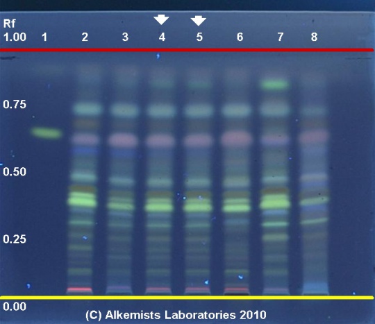
Ganoderma lucidum HPTLC ID - 10% Methanolic Sulfuric Acid - UV 365 nm
Reishi (fruiting body) (Ganoderma lucidum)
Lane Assignments Lanes, from left to right (Track, Volume, Sample):
- 3 μL Ursolic Acid ~ 0.1% Methanol
- 3 μL Ganoderma lucidum-1 (fruiting body)
- 3 μL Ganoderma lucidum-2 (fruiting body)
- 3 μL Ganoderma lucidum-3 (fruiting body)
- 3 μL Ganoderma lucidum-3 (fruiting body)
- 3 μL Ganoderma lucidum-4 (fruiting body)
- 3 μL Ganoderma lucidum-5 (fruiting body)
- 3 μL Ganoderma lucidum-6 (fruiting body)
Reference materials used here have been authenticated by macroscopic, microscopic &/or TLC studies according to the reference source cited below held at Alkemists Laboratories, Costa Mesa, CA.
Stationary Phase Silica gel 60, F254, 10 x 10 cm HPTLC plates
Mobile Phase Dichloromethane : Methanol [9/1]
Sample Preparation Method 0.3g+3mL 70% grain EtOH sonicate/heat @ 50° C ~ 1/2 hr
Detection Method 10% Methanolic H2SO4 -> UV 365 nm
Reference see American Herbal Pharmacopoeia & Therapeutic Compendium
Source: Elan M. Sudberg, Alkemist Laboratories [2]
|
Other Points of Interest
Cite error: <ref> tags exist, but no <references/> tag was found

