Revision as of 22:13, 15 March 2013 by
JKim (Talk)
Introduction
Macroscopic Entries
Microscopic Entries
| Fragment of the bark showing dispersal of medullary rays observed at 400x with Acidified Chloral Hydrate Glycerol Solution.cellular structures identified in this botanical specimen is the fragment of the bark showing dispersal of medullary rays and the bark fiber showing calcium oxalate prisms spaced by medullary rays when observed at 400x with Acidified Chloral Hydrate Glycerol Solution.
Source: Elan M. Sudberg, Alkemist Laboratories [1]
|
|
|
|
HPTLC Entries
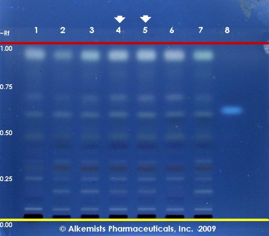
Handroanthus impetiginosus HPTLC ID - 10% Ethanolic H2SO4 ->115° C 15 min -> UV 365 nm
Pau D’Arco (bark) (Handroanthus impetiginosus)
Lane Assignments Lanes, from left to right (Track, Volume, Sample):
- 3 μL Handroanthus impetiginosus-1 (bark)
- 3 μL Handroanthus impetiginosus-2 (bark)
- 3 μL Handroanthus impetiginosus-3 (bark)
- 3 μL Handroanthus impetiginosus-4 (bark)
- 3 μL Handroanthus impetiginosus-4 (bark)
- 3 μL Handroanthus impetiginosus-5 (bark)
- 3 μL Handroanthus impetiginosus-6 (bark)
- 1 μL Artemisinin ~0.1% in CH3OH
Reference materials used here have been authenticated by macroscopic, microscopic &/or TLC studies according to the reference source cited below held at Alkemists Laboratories, Costa Mesa, CA.
Stationary Phase Silica gel 60, F254, 10 x 10 cm HPTLC plates
Mobile Phase toluene: ethyl acetate: formic acid [7.5/2.5/0.5]
Sample Preparation Method 0.3 g + 3 ml CH3OH sonicated + heated @ 50° C ~ 1 hr.
Detection Method 10% Ethanolic H2SO4 ->115° C 15 min -> UV 365 nm
Reference see Plant Drug Analysis, Wagner, H., 1996
Source: Elan M. Sudberg, Alkemist Laboratories [2]
|
Other Points of Interest
Cite error: <ref> tags exist, but no <references/> tag was found

