Hypericum perforatum (flowering tops)

From AHPA Botanical Identity References Compendium
Revision as of 04:43, 22 March 2014 by Staffer (Talk | contribs)

Jump to: navigation, search

Contents

Nomenclature

Hypericum perforatum L.   Clusiaceae  
Standardized common name (English): St. John's wort

Botanical Voucher Specimen

bottomright bottomright bottomright bottomright

St. John's Wort Tropicos 61762.jpg
Source: MOBOT, Tropicos.org.[1]

Hypericum perforatum LF00509MMB A0153.jpg
Source: Botanical Voucher Specimen Library, Alkemists Laboratories[2]

Hypericum perforatum LF20105BMX1 A0155.jpg
Source: Botanical Voucher Specimen Library, Alkemists Laboratories[3]

Hypericum perforatum LF19505JD A0154.jpg
Source: Botanical Voucher Specimen Library, Alkemists Laboratories[4]

Organoleptic Characteristics

Color: Stem greenish-yellow to brownish-yellow; leaves greenish-grey; flowers, green sepals, golden-yellow petals

Aroma/Odor: Weak, aromatic, balsamic Flavor/Taste: Bitter, acrid

Source: WHO monographs on selected medicinal plants, Vol.2, World Health Organization, Geneva, 1999 [5]

Macroscopic Characteristics

"...Stem glabrous greenish-yellow to brownish-yellow branching, 2-winged, cylindrical with 2 equidistant longitudinal bands. Leaves glabrous, generally sessile, opposite, greenish-grey, oval, 8–35mm long, with entire margins; laminal margin often more or less revolute-marginated. Brown-black glandular dots sometimes present along the edges; numerous pellucid glands on the entire surface. Flowers, 2 cm in diameter, regular, forming a broadly paniculate, compound cymose inflorescence at top of stem, composed of: 5 green, lanceolate sepals, containing punctiform, black glandular dots on the edges; 5 golden-yellow petals, with numerous glandular dots along margins; and 3 staminal blades, each divided into multiple golden-yellow stamens. Anthers with single, terminal, dark pigment dot. Ovary elongated and conical, parietal placentation, carries 3 styles."

Source: WHO monographs on selected medicinal plants, Vol.2, World Health Organization, Geneva, 1999 [6]

bottomright bottomright bottomright bottomright

Hypericum perforatum 1 RU.jpg
Hypericum perforatum growing in the field
Source: Roy Upton, American Herbal Pharmacopoeia®[7]

Hypericum blossom stereo 2.jpg
Hypericum perforatum flower close-up. Note the black dots on the petals' margin only.
Source: American Herbal Pharmacopoeia®[8]

Hypericum perforatum leaves stereo 1.JPG
Hypericum perforatum leaves with the characteristic black dots on the margin and translucent dots on the leaf body.
Source: American Herbal Pharmacopoeia®[9]

Hypericum perforatum stem stereo 2.JPG
Hypericum perforatum stem showing opposite ridges, a defining characteristic of the species.
Source: American Herbal Pharmacopoeia®[10]

Microscopic Characteristics

Transverse section: "...stem circular and presents 2 lateral edges corresponding to the 2 longitudinal bands. From the exterior inwards are seen: epidermal layer formed of large polygonal cells; continuous collenchymal layer, slightly more developed at the 2 lateral edges; a cortical parenchyma containing crystals of calcium oxalate in the shape of a sea urchin; a ring of continuous phloem, distinct from the xylem, which consists of large vessels and a lignified parenchyma with a visible cambium; and a lacunose medullary parenchyma. Secretory pockets, almost invisible, rarely present in the endoderm. Upper surface of leaf section shows polygonal cells with sinuous, slightly beaded, anticlinal walls; cells of lower surface smaller, anticlinal walls more wavy with frequent paracytic, sometimes anomocytic, stomata; smooth cuticle, thicker on upper surface; straight-walled, elongated epidermal cells of veins occasionally beaded. Dorsoventral surface of leaf consists of a single palisade layer and large oil glands. Midrib shows single, collateral bundle with small area of lignified xylem. Microscopic characteristics of the sepal resemble those of the leaf. Petal narrow, elongated, thin-walled, epidermal cells with straight anticlinal walls on outer surface and wavy on inner surface. Stamen lignified fibrous layer of anther wall; elongated, thin-walled cells of filament with striated cuticle. Pollen grains spherical or elliptical, 20–28mm in diameter, with 3 germinal pores and smooth exine."

Powder: "...Yellowish-green or brownish-green. Leaf fragments abundant, most containing large characteristic hypericin oil glands with brown to red contents. Fragments of leaf epidermis, the adaxial side with thick-walled punctate, slightly sinuate cells, and abaxial side with sinuate cells and paracytic stomata; mesophyll fragments with large secretory pockets which are spherical, bright, containing strongly refractive oil droplets; fragments of palisade parenchyma; stem fragments with reticulate spiral vessels, areolate punctation, long fibres with thick walls, ligneous parenchyma, and small number of thick-walled, characteristically punctate medullary cells; fragments of petals made of elongated rectangular cells with irregular nodulous thickenings, containing numerous yellow droplets and large, round to oval secretory pockets; fragments of anthers; pollen grains 20–28mm in diameter, smooth spherical or elliptical with 3 germinal pores; clusters of calcium oxalate crystals."

Source: WHO monographs on selected medicinal plants, Vol.2, World Health Organization, Geneva, 1999 [11]

bottomright bottomright bottomright bottomright

Hypericum perforatum L. -Clusiaceae--1.jpg
Oil glands with red pigment of Hypericum perforatum viewed at 400x with Acidified Chloral Hydrate Solution.
Source: Elan M. Sudberg, Alkemist Laboratories[12]

Hypericum perforatum L. -Clusiaceae--2.jpg
Three pored pollen grain showing smooth and faintly warted exine of Hypericum perforatum viewed at 400x with Acidified Chloral Hydrate Solution.
Source: Elan M. Sudberg, Alkemist Laboratories[13]

St johns wort secretory cell phytolab.jpg
St. John's Wort, Hyperici herba secretory cell
Source: Claudia Borst, PhytoLab[14]

St johns wort pollen grain phytolab.jpg
St. John's Wort, Hyperici herba pollen grain
Source: Claudia Borst, PhytoLab[15]

bottomright bottomright

St johns wort hypericin gland phytolab.jpg
St. John's Wort, Hyperici herba hypericin gland
Source: Claudia Borst, PhytoLab[16]

St johns wort tracheidal vessels with pitted walls phytolab.jpg
St. John's Wort, Hyperici herba tracheidal vessels with pitted walls
Source: Claudia Borst, PhytoLab[17]


High Performance Liquid Chromatographic Identification

Indena - Logo.png
(thumbnail)
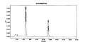
HYPERICUM PERFORATUM DRY EXTRACT - Total hypericins HPLC assay
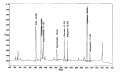
'Method for Hypericins

Column: 15-cm x 4.6 mm, 5-um, Waters Atlantis C18

Mobile Phase: A mix of 39 volumes of ethyl acetate, 41 volumes of a 15.6 g/L solution of sodium dihydrogen phosphate adjusted to pH 2 with phosphoric acid and 160 volumes of methanol.

Elution: Isocratic

Column Temperature: 40°C

Flow rate: 1.0 mL/min

Detection: Vis, 590 nm

Injection volume: 20 uL

Method for Hyperforins and Flavonoids

Column: 15-cm x 4.6 mm, 3-um, YMC-Pack ODS-A

Mobile Phase: Water, containing 0.3 % (v/v) phosphoric acid 85% (Solution A) and acetonitrile containing 0.3 % (v/v) phosphoric acid 85% (Solution B)

Elution: Gradient, see table below

Column Temperature: 25°C

Detection: UV, 360 nm, then at 275 nm after the elution of biapigenin (about 22 min)

Injection volume: 10 uL

Source: Indena S.p.A. [18]


Table: Gradient program

Time (min) Solution A (%) Solution B (%) Flow Rate (mL/min)
0-8 82 18 0.8
8-18 82-47 18-53 0.8
18-18.1 47-3 53-97 0.8
18.1-19 3 97 1.2
19-29 3 97 1.2
29-30 82 18 1.2
30-31 82 18 0.8
31-35 82 18 0.8

High Performance Thin Layer Chromatographic Identification

HPTLC-assoc-Logo-farbig-Text-schwarz-300x47.png
(thumbnail)
Hypericum perforatum HPTLC, UV 366 nm

St. John's wort (herb) (Hypericum perforatum)

Lane Assignments Lanes, from left to right (Track, Volume, Sample):

  1. 2 μL Rutin, Hyperoside (increasing Rf)
  2. 2 μL Chlorogenic acid, Hypericin, Pseudohypericin (increasing Rf)
  3. 4 μL St. John's wort herb 1
  4. 4 μL St. John's wort herb 2
  5. 4 μL St. John's wort herb 3
  6. 4 μL St. John's wort herb 4
  7. 4 μL St. John's wort herb 5
  8. 4 μL Mountain St. John's wort herb
  9. 4 μL Hairy St. John's wort herb
  10. 4 μL Wavy St. John's wort herb 

Other Notes
Reference Standard Solution: 1.0 mg/mL rutin in methanol, 0.1 mg/mL hyperoside, 0.1 mg/mL hypericin and 0.1 mg/mL pseudohypericin (sonicate for 10 min, centrifuge, and use the supernatant), and 1.0 mg/mL chlorogenic acid in methanol (optional).

Reference Sample Preparations: Sonicate 0.5 g of powdered sample in 5 mL of methanol at 60°C, centrifuge or filter the solution, and use the supernatant / filtrate.

Stationary Phase: HPTLC, Silica gel 60 F254

Mobile Phase: Ethyl acetate, dichloromethane, water, formic acid, acetic acid (100:25:11:10:10)

Development: Saturate chamber for 20 minutes; developing distance 70 mm from lower edge of the plate; relative humidity 33%, temperature 25°.

Derivatization reagent A: Natural Products Reagent (NP)– 5 mg/mL 2-aminoethyl diphenylborinate in ethyl acetate in a glass bottle.

Derivatization reagent B: PEG Reagent– 50 mg/mL polyethylene glycol 400 (macrogol) in dichloromethane in a glass bottle.

Detection: Heat plate at 100°C for 3 min, dip (time 0, speed 5) in Derivatization reagent A, dry, dip (time 0, speed 5) in Derivatization reagent B, and examine under UV light at 366 nm.

System suitability: Rutin: orange fluorescent zone in the lower one-third of the plate at about Rf 0.1. Hyperoside: orange fluorescent zone in the lower one-third of the plate at about Rf 0.25.

Procedure:

Reference Standard Solutions, Stationary Phase, Mobile Phase, Development, Derivatization reagent A, Derivatization reagent B, and Detection, as described above.

Test Sample Preparation: Prepare test sample as described under Reference Sample Preparations and apply 4 uL.

Identification: Compare Test Sample Preparation chromatogram with chromatograms of Reference Sample Preparations. The Test Sample Preparation chromatogram is similar to that of the Reference Sample Preparations chromatograms. Additional weak zones may be present.

The Test Sample Preparation chromatogram exhibits fluorescent zones corresponding to the zones due to rutin, hyperoside, hypericin, and pseudohypericin in the Reference Standard Solution chromatograms. Between the zones due to hyperoside and hypericin there are several orange fluorescent zones.

Related Species: A very faint zone at the Rf corresponding to rutin indicating Hypericum montanum (mountain St. John’s Wort), Hypericum hirsutum (hairy St. John’s Wort), or Hypericum undulatum (wavy St. John’s Wort). A blue fluorescent zone detected below and well resolved from the hyperoside zone indicating Hypericum montanum. Two yellow zones detected around one-third of the chromatogram indicating Hypericum hirsutum. An orange zone detected at Rf 0.47 indicating Hypericum undulatum.

Note: Images presented in this entry are examples and are not intended to be used as a bases for setting specifications for quality control purposes.


Source: HPTLC Association [19]


AP-LOGO-Laboratories Crop - Copy.jpg
(thumbnail)
Hypericum perforatum HPTLC ID - Natural Product Reagent + PEG UV 365 nm

St. John's Wort (herb) (Hypericum perforatum)

Lane Assignments Lanes, from left to right (Track, Volume, Sample):

  1. 3 μL Hypericin ~ 0.1% in Methanol
  2. 3 μL Hypericum perforatum-1 (herb)
  3. 3 μL Hypericum perforatum-2 (herb)
  4. 3 μL Hypericum perforatum-3 (herb)
  5. 3 μL Hypericum perforatum-3 (herb)
  6. 3 μL Hypericum perforatum-4 (herb)
  7. 3 μL Hypericum perforatum-5 (herb)
  8. 1 μL Rutin, Caffeic Acid, Hyperoside, Chlorogenic Acid ~ 0.1% in Methanol

Reference materials used here have been authenticated by macroscopic, microscopic &/or TLC studies according to the reference source cited below held at Alkemists Laboratories, Costa Mesa, CA. 

Stationary Phase Silica gel 60, F254, 10 x 10 cm HPTLC plates 

Mobile Phase ethyl acetate: Acetic acid: HCOOH: H2O [10/1.1/1.1/2.4] 

Sample Preparation Method 0.3 g + 3 ml 70% grain EtOH sonicated + heated @ 50° C ~ 1 hr 

Detection Method Natural Product Reagent + PEG -> UV 365 nm 

Reference see British Pharmacopoeia, 2003


Source: Elan M. Sudberg, Alkemist Laboratories [20]

Supplementary Information

Sources

  1. MOBOT, Tropicos.org. http://www.tropicos.org/Image/61762
  2. Botanical Voucher Specimen Library, Alkemists Laboratories http://www.alkemist.com
  3. Botanical Voucher Specimen Library, Alkemists Laboratories http://www.alkemist.com
  4. Botanical Voucher Specimen Library, Alkemists Laboratories http://www.alkemist.com
  5. WHO monographs on selected medicinal plants, Vol.2, World Health Organization, Geneva, 1999
  6. WHO monographs on selected medicinal plants, Vol.2, World Health Organization, Geneva, 1999
  7. Roy Upton, American Herbal Pharmacopoeia® http://www.herbal-ahp.org/
  8. American Herbal Pharmacopoeia® http://www.herbal-ahp.org/
  9. American Herbal Pharmacopoeia® http://www.herbal-ahp.org/
  10. American Herbal Pharmacopoeia® http://www.herbal-ahp.org/
  11. WHO monographs on selected medicinal plants, Vol.2, World Health Organization, Geneva, 1999
  12. Elan M. Sudberg, Alkemist Laboratories http://www.alkemist.com
  13. Elan M. Sudberg, Alkemist Laboratories http://www.alkemist.com
  14. Claudia Borst, PhytoLab http://www.phytolab.com/en.html
  15. Claudia Borst, PhytoLab http://www.phytolab.com/en.html}
  16. Claudia Borst, PhytoLab http://www.phytolab.com/en.html
  17. Claudia Borst, PhytoLab http://www.phytolab.com/en.html
  18. Indena S.p.A. http://www.indena.com/
  19. HPTLC Association http://www.hptlc-association.org/
  20. Elan M. Sudberg, Alkemist Laboratories http://www.alkemist.com
Personal tools
MediaWiki Appliance - Powered by TurnKey Linux