Lavandula angustifolia (flower)
(add askbox) |
(add USD 1918 organoleptic information) |
||
| Line 13: | Line 13: | ||
=Botanical Voucher Specimen= | =Botanical Voucher Specimen= | ||
=Organoleptic Characteristics= | =Organoleptic Characteristics= | ||
| − | + | {| border=1 | |
| + | | | ||
| + | {{Organolepsy | source=United States Dispensatory (1918) | ||
| + | | description=Lavender flowers have a strong fragrant odor, and an aromatic, warm, bitterish taste. They retain their fragrance long after drying.}} | ||
| + | |} | ||
| + | |||
=Macroscopic Characteristics= | =Macroscopic Characteristics= | ||
=Microscopic Characteristics= | =Microscopic Characteristics= | ||
Revision as of 14:18, 31 March 2015
Contents |
Nomenclature
Lavandula angustifolia Mill. Lamiaceae
Syn. Lavandula officinalis Chaix.; Lavandula spica L.; Lavandula vera DC.
Standardized common name (English): English lavender
Botanical Voucher Specimen
Organoleptic Characteristics
|
Macroscopic Characteristics
Microscopic Characteristics
High Performance Thin Layer Chromatographic Identification
|
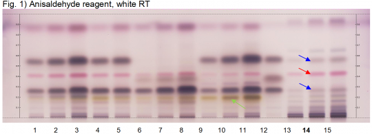
Lavender flower (flower) (Lavandula angustifolia) Lane Assignments Lanes, from left to right (Track, Volume, Sample):
Reference Sample(s) Reference: Dissolve 5 µL of linalool in 1 mL of toluene; Dissolve 5 µL of linalyl acetate in 1mL of toluene; Optional: Dissolve 10 µL of cineole in 1 mL of toluene. Stationary Phase Stationary phase, i.e. Silica gel 60, F254 Mobile Phase Toluene, ethyl acetate 95:5 (v/v) Sample Preparation Method Sample: Mix 500 mg of powdered sample with 5 mL of toluene and sonicate for 10 minutes, centrifuge or filter the solutions and use the supernatants / filtrates as test solutions. Derivatization reagent: Anisaldehyde reagent, Preparation: 170 mL of ice-cooled methanol are mixed with 20 mL of acetic acid, 10 mL of sulfuric acid and 1 mL of anisaldehyde, Use: Dip (time 0, speed 5), heat at 100°C for 5 min. Detection Method Saturated chamber; developing distance 70 mm from lower edge; relative humidity 33%. Other Notes Images presented in this entry are examples and are not intended to be used as basis for setting specifications for quality control purposes. System suitability test: Linalyl acetate: violet zone at Rf ~ 0.57; Linalool: violet zone at Rf ~ 0.27. Identification: Compare result with reference images. The fingerprint of the test solution is similar to that of the corresponding botanical reference sample. Additional weak zones may be present. The chromatogram of the test solution shows a violet zone at Rf ~ 0.27 corresponding to reference linalool and a violet zone at Rf ~ 0.57 corresponding to linalyl acetate (blue arrows). Between these zones there is a reddish violet zone at Rf ~ 0.42 (red arrow). There is a grey zone at Rf ~ 0.66 above linalyl acetate. Test for other species: No yellow zone is seen between the application position and reference linalool (green arrow) (Lavender oil, Lavandin, and Spike lavender oil). Lavender oil, Lavandin, and Spike lavender oil all show a more intense violet zone at the position of linalool. Lavender oil and Lavandin also show a more intense violet zone at the position of linalyl acetate.
|
|
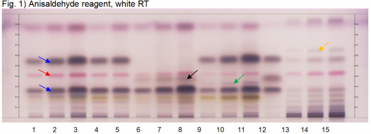
Lavender oil (flower) (Lavandula angustifolia) Lane Assignments Lanes, from left to right (Track, Volume, Sample):
Reference Sample(s) Reference: Dissolve 5 µL of linalool in 1 mL of toluene; Dissolve 5 µL of linalyl acetate in 1mL of toluene; Optional: Dissolve 10 µL of cineole in 1 mL of toluene. Stationary Phase Stationary phase, i.e. Silica gel 60, F254 Mobile Phase Toluene, ethyl acetate 95:5 (v/v) Sample Preparation Method Sample: Mix 500 mg of powdered sample with 5 mL of toluene and sonicate for 10 minutes, centrifuge or filter the solutions and use the supernatants / filtrates as test solutions. Derivatization reagent: Anisaldehyde reagent, Preparation: 170 mL of ice-cooled methanol are mixed with 20 mL of acetic acid, 10 mL of sulfuric acid and 1 mL of anisaldehyde, Use: Dip (time 0, speed 5), heat at 100°C for 5 min. Detection Method - Saturated chamber; developing distance 70 mm from lower edge; relative humidity 33%. Other Notes Images presented in this entry are examples and are not intended to be used as basis for setting specifications for quality control purposes. System suitability test: Linalyl acetate: violet zone at Rf ~ 0.57; Linalool: violet zone at Rf ~ 0.27. Identification: Compare result with reference images. The fingerprint of the test solution is similar to that of the corresponding botanical reference sample. Additional weak zones may be present. The chromatogram of the test solution shows an intense violet zone at Rf ~ 0.27 corresponding to reference linalool and an intense violet zone at Rf ~ 0.57 corresponding to linalyl acetate (blue arrows). Between these zones there is a violet-red zone at Rf ~ 0.42 (red arrow). Between the red zone and the zone due to linalool there may be a faint violet zone (cineole). Test for other species: The chromatogram of Spike lavender oil does not show a zone at the position of linalyl acetate. There is a grey zone at the position of cineole (black arrow). The chromatogram of Lavandin shows a narrow violet zone between linalool and cineole (green arrow). Dried Lavender flower shows less intense violet zones due to linalool and linalyl acetate and an additional grey zone above linalyl acetate (orange arrow).
|
Supplementary Information
Sources
- ↑ United States Dispensatory (1918)
- ↑ HPTLC Association http://www.hptlc-association.org/
- ↑ HPTLC Association http://www.hptlc-association.org/

