Matricaria recutita (flower)
Contents |
Nomenclature
Botanical Voucher Specimen
Organoleptic Characteristics
|
Macroscopic Descriptions
|
Microscopic Characteristics
|
High Performance Thin Layer Chromatographic Identification
|
Chamomile (flower) (Matricaria recutita) Lane Assignments Lanes, from left to right (Track, Volume, Sample):
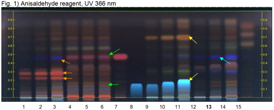
Reference Sample(s) Reference: Dissolve 1.5 mg of apigenin in 5 mL of methanol. Dissolve 1 mg of parthenolide in 5 mL of methanol. Stationary Phase Stationary phase, i.e. Silica gel 60, F254 Mobile Phase Ethyl acetate, cyclohexane 1:1 (v/v) Sample Preparation Method Sample: Mix 1 g of powdered sample with 10 mL of methanol and sonicate for 10 minutes, then centrifuge or filter the solutions and use the supernatants / filtrates as test solutions. Dissolve 10 µL of the essential oil in 1 mL of toluene. Derivatization reagent: Anisaldehyde reagent; Preparation: 170 mL of ice-cooled methanol are mixed with 20 mL of acetic acid, 10 mL of sulfuric acid and 1 mL of anisaldehyde; Use: Dip (time 0, speed 5), heat at 100°C for 4 min. Detection Method Saturated chamber; developing distance 70 mm from lower edge; relative humidity 33% Other Notes Images presented in this entry are examples and are not intended to be used as basis for setting specifications for quality control purposes. System suitability test (UV 366 nm): Apigenin: blue zone at Rf ~ 0.20; Parthenolide: pink zone at Rf ~ 0.48 Identification: Compare result with reference images. The fingerprint of the test solution is similar to that of the corresponding botanical reference sample. Additional weak zones may be present. Under UV 366 nm the chromatogram of the test solution shows a dark blue zone at Rf ~ 0.46 (blue arrow; not visible under white light) a brown zone between Rf ~ 0.30 and 0.40 and another weak blue zone at Rf ~ 0.30. At the position of apigenin reference substance (Rf ~ 0.20) two brown zones are present (violet under white light). Test for adulteration: Under UV 366 nm no pink zone (violet zone under white RT) at Rf ~ 0.48 corresponding to reference substance parthenolide is seen and there are no brown zones at the position of apigenin at Rf ~ 0.20 (green arrows, Feverfew flower). Under UV 366 nm there are no intense red zones (violet zones under white RT) between Rf ~ 0.20 and 0.30. No blue zone is seen at the position of parthenolide (orange arrows; Feverfew flower from Mexico). Under UV 366 nm no blue zone at the position of apigenin (Rf ~ 0.20) and no orange zone at Rf ~ 0.70 (purple under white light) is seen (yellow arrows; Roman Chamomile flower). Chamomile flower oil does not show any zones below Rf ~ 0.60. Source: HPTLC Association [9] |
Supplementary Information
Sources
- ↑ Steven Yeager, Mountain Rose Herbs http://www.MountainRoseHerbs.com
- ↑ Schneider, A. (1921) The Microanalysis of Powdered Vegetable Drugs, 2nd ed.
- ↑ Steven Yeager, Mountain Rose Herbs http://www.MountainRoseHerbs.com
- ↑ Schneider, A. (1921) The Microanalysis of Powdered Vegetable Drugs, 2nd ed.
- ↑ Amy Brush, Traditional Medicinals http://www.traditionalmedicinals.com
- ↑ WWWWWWWWWWWWWWWW
- ↑ WWWWWWWWWWWWWWWW
- ↑ Amy Brush, Traditional Medicinals http://www.traditionalmedicinals.com
- ↑ HPTLC Association http://www.hptlc-association.org/


