Melissa officinalis (leaf)
Contents |
Nomenclature
Melissa officinalis L. Lamiaceae
Standardized common name (English): lemon balm
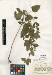
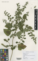
Botanical Voucher Specimen
 |
|
|
|
|
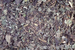
Organoleptic Characteristics
|
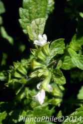
Macroscopic Characteristics
|
Microscopic Characteristics
High Performance Thin Layer Chromatographic Identification
|
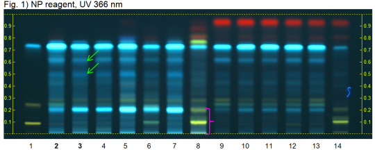
Melissa leaf dry extract (leaf) (Melissa officinalis) Lane Assignments Lanes, from left to right (Track, Volume, Sample):
Reference Sample(s) Reference: Dissolve 1 mg of hyperoside, 1 mg of rutin and 5 mg of rosmarinic acid individually in 10 mL of methanol. Stationary Phase Stationary phase, i.e. Silica gel 60, F254 Mobile Phase Formic acid, water, ethyl acetate 1:1:15 (v/v/v) Sample Preparation Method Sample: Mix 0.2 g of extract with 5 mL of methanol and sonicate for 5 minutes, then centrifuge or filter the solutions and use the supernatants / filtrates as test solutions. Optional: Mix 0.5 g of dried Melissa leaf with 5 mL methanol and proceed as above. Derivatization reagent: NP reagent, Preparation: 1 g of natural products reagent in 200 mL of ethyl acetate; 2.) PEG reagent, Preparation: 10 g of polyethylene glycol 400 in 200 mL of methylene chloride, Use: Heat plate for 3 min at 100°C, dip (time 0, speed 5) in NP reagent, dry and dip (time 0, speed 5) in PEG reagent, dry in air. Detection Method Saturated chamber; developing distance 70 mm from lower edge; relative humidity 33% Other Notes Images presented in this entry are examples and are not intended to be used as basis for setting specifications for quality control purposes. System suitability test: Rutin: yellow fluorescent zone Rf ~ 0.09; Hyperoside: yellow fluorescent zone at Rf ~ 0.25; Rosmarinic acid: light blue fluorescent zone at Rf ~ 0.74. Identification: Compare result with reference images. The fingerprint of the test solution is similar to that of the corresponding botanical reference sample. Additional weak zones may be present. The chromatogram of the test solution shows a light blue fluorescent zone just below the position of reference hyperoside. At the position of rosmarinic acid there is an intense light blue fluorescent zone. Between this zone and reference hyperoside there are two bluoe fluorescent zones (green arrows). Test for other species: No yellow zones are seen below the position of reference hyperoside (pink arrow, Peppermint dry extract).
|
Supplementary Information
Sources
- ↑ MOBOT, Tropicos.org http://www.tropicos.org/Image/100007389
- ↑ Royal Botanic Gardens, Kew. http://specimens.kew.org/herbarium/K000914345
- ↑ Culbreth, D. (1917) A Manual of Materia Media and Pharmacology, 6th ed.
- ↑ American Medicinal Plants of Commercial Importance (1930)
- ↑ Culbreth, D. (1917) A Manual of Materia Media and Pharmacology, 6th ed.
- ↑ American Medicinal Plants of Commercial Importance (1930)
- ↑ PlantaPhile http://plantaphile.com/
- ↑ PlantaPhile http://plantaphile.com/
- ↑ HPTLC Association http://www.hptlc-association.org/
