Piper methysticum (rhizome)
Contents |
Nomenclature
Piper methysticum G. Forst. Piperaceae
Standardized common name (English): kava
Botanical Voucher Specimen
Organoleptic Characteristics
Macroscopic Characteristics
Microscopic Characteristics
High Performance Thin Layer Chromatographic Identification
|
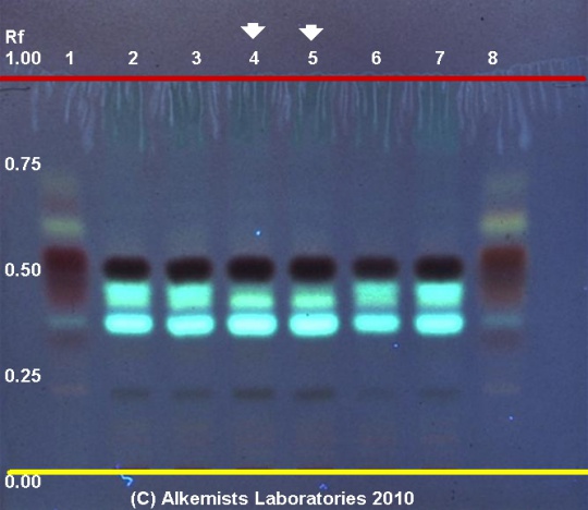
Kava (rhizome) (Piper methysticum) Lane Assignments Lanes, from left to right (Track, Volume, Sample):
Reference materials used here have been authenticated by macroscopic, microscopic &/or TLC studies according to the reference source cited below held at Alkemists Laboratories, Costa Mesa, CA. Stationary Phase Silica gel 60, F254, 10 x 10 cm HPTLC plates Mobile Phase H2O: acetonitrile: CH3OH: AcCOOH: >Reverse Phase!!!! [4/3/3/0.01/] Sample Preparation Method 0.3g+3mL 70% grain EtOH sonicate/heat @ 50° C ~ 1/2 hr Detection Method Vanillin/H2SO4 Reagent -> 110° C 5 min -> UV 365 nm Reference see Plant Drug Analysis, Wagner, H., 1996
|
|
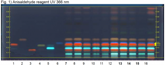
Kava (rhizome) (Piper methysticum) Lane Assignments Lanes, from left to right (Track, Volume, Sample):
Reference Sample(s) Reference: Dissolve 1 mg of kavain in 2 mL of toluene. Dissolve 1 mg of desmethoxyyangonin in 2 mL of toluene. Optional: dissolve 1 mg of dihydrokavain, 1 mg of methysticin, 1 mg of dihydromethysticin, and 1 mg of yangonin each in 2 mL of toluene. Stationary Phase Stationary phase, i.e. Silica gel 60, F254, caffeine impregnated Mobile Phase tert-Butyl methyl ether, n-hexane 7:3 (v/v) Sample Preparation Method Sample: Mix 1 g of powdered sample with 10 mL of methanol and sonicate for 10 minutes, then centrifuge or filter the solutions and use the supernatants / filtrates as test solutions. Derivatization reagent: Anisaldehyde reagent; Preparation: 170 mL of ice-cooled methanol are mixed with 20 mL of acetic acid, 10 mL of sulfuric acid and 1 mL of anisaldehyde; Use: Dip (time 0, speed 5), heat at 100°C for 4 min. Detection Method Unsaturated chamber; developing distance 70 mm from lower edge; relative humidity 33% Other Notes Images presented in this entry are examples and are not intended to be used as basis for setting specifications for quality control purposes. System suitability test (UV 366 nm): Kavain: reddish zone at Rf ~ 0.27; Desmethoxyyangonin: blue zone at Rf ~ 0.30 Identification: Compare result with reference images. The fingerprint of the test solution is similar to that of the corresponding botanical reference sample. Additional weak zones may be present. Under UV 366 nm the chromatogram of the test solution shows a green zone (grey zone under white RT) at Rf ~ 0.08, a red zone (purple zone under white RT) at Rf ~ 0.16, another green zone (grayish and weak zone under white RT) at Rf ~ 0.19, another red zone (red and intense zone under white RT) at Rf ~ 0.27 corresponding to reference substance kavain, just above it a faint blue zone at Rf ~ 0.30 corresponding to reference substance desmethoxyyangonin (yellow arrows), and a brown zone (green zone under white RT) at Rf ~ 0.35. Under white light there are several yellow and reddish zones in the upper part of the chromatogram. Plate preparation: Dissolve 8 g of caffeine in 200 mL of dichloromethane. Use: Dip (time 0, speed 5) plate, dry at room temperature for 5 minutes, then heat at 80°C for 5 minutes. Source: HPTLC Association [2] |
Supplementary Information
Sources
- ↑ Elan M. Sudberg, Alkemist Laboratories http://www.alkemist.com
- ↑ HPTLC Association http://www.hptlc-association.org/


