Viola spp. (aerial parts)
Contents |
Nomenclature
Botanical Voucher Specimen
Organoleptic Characteristics
Macroscopic Characteristics
Microscopic Characteristics
High Performance Thin Layer Chromatographic Identification
|
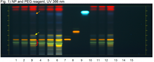
Wild pansy flowering (aerial parts) (Viola arvensis, V. tricolor) Lane Assignments Lanes, from left to right (Track, Volume, Sample):
Reference Sample(s) Reference: Individually dissolve 2.5 mg of rutin, 1.0 mg of caffeic acid, and 2.5 mg of hyperoside each in 10 mL of methanol. Stationary Phase Stationary phase, i.e. Silica gel 60, F254 Mobile Phase Formic acid, acetic acid, water, ethyl acetate 11:11:27:100 (v/v/v/v) Sample Preparation Method Sample: Mix 2 g of powdered sample with 10 mL of methanol and sonicate for 10 minutes, then centrifuge or filter the solutions and use the supernatants / filtrates as test solutions. Derivatization reagent: 1.) NP reagent, Preparation: 1 g of natural products reagent in 200 mL of ethyl acetate; 2.) PEG reagent, Preparation: 10 g of polyethylene glycol 400 in 200 mL of dichloromethane, Use: Heat plate for 3 min at 100°C, dip (time 0, speed 5) in NP reagent, dry and dip (time 0, speed 5) in PEG reagent Detection Method Saturated chamber; developing distance 70 mm from lower edge; relative humidity 33% Other Notes Images presented in this entry are examples and are not intended to be used as basis for setting specifications for quality control purposes. System suitability test: Caffeic acid: blue fluorescent zone at Rf ~ 0.87. Rutin: orange fluorescent zone at Rf ~ 0.32. Identification: Compare result with reference images. The fingerprint of the test solution is similar to that of the corresponding botanical reference sample. Additional weak zones may be present. The chromatogram of the test solution shows a blue fluorescent zone just below the zone due to reference caffeic acid (orange arrow). A yellow fluorescent zone is seen at Rf ~ 0.40 below the position of reference hyperoside (yellow arrow). Below it there is an intense yellowish orange fluorescent zone corresponding to reference rutin. Below the zone due to rutin there are three yellowish-green fluorescent zones (red arrow).
|
Supplementary Information
Sources
- ↑ HPTLC Association http://www.hptlc-association.org/
